
ResearchMedical imaging & computer vision
Digital Microscopy Image Analysis
Automated detection, quantification and explainable classification of structures in microscopy and histopathology images.
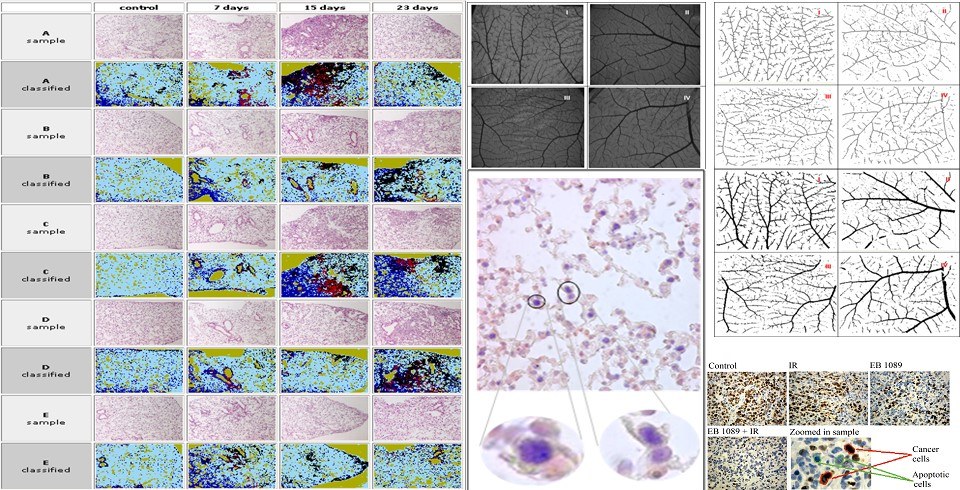
Microscopy and histopathology images contain thousands of cells and tissue structures that are slow and subjective to assess by eye. We develop tools that detect, quantify and classify these structures automatically.
Applications include the quantification of fibrotic tissue, the characterisation of breast cancer and kidney biopsies, and the classification of histopathology images with ensembles of convolutional neural networks, together with methods that make their decisions explainable to pathologists.
- Fibrosis and tissue quantification
- Cancer histopathology classification
- Explainable deep learning for pathology
Related publications
Selected work from the lab in this area.
FAST: A Dual-tier Few-Shot Learning Paradigm for Whole Slide Image Classification
In Advances in Neural Information Processing Systems 37. Neural Information Processing Systems Foundation, Inc. (NeurIPS), pp. 105090–105113.
On the evaluation of deep learning interpretability methods for medical images under the scope of faithfulness
Computer Methods and Programs in Biomedicine, 253, p. 108238.
Improving explainability results of convolutional neural networks in microscopy images
Neural Computing and Applications, 35(29), pp. 21535–21553.
Ensembling to Leverage the Interpretability of Medical Image Analysis Systems
IEEE Access, 11, pp. 76437–76447.
Explainable computer vision analysis for embryo selection on blastocyst images
In 2022 IEEE-EMBS International Conference on Biomedical and Health Informatics (BHI). IEEE, pp. 1–4.
Image Analysis in Digital Pathology Utilizing Machine Learning and Deep Neural Networks
Journal of Personalized Medicine, 12(9), p. 1444.
Ensembling EfficientNets for the Classification and Interpretation of Histopathology Images
Algorithms, 14(10), p. 278.
Creating Visual Vocabularies for The Retrieval And Classification of Histopathology Images
In 2019 41st Annual International Conference of the IEEE Engineering in Medicine and Biology Society (EMBC). IEEE, pp. 7036–7039.
Content Based Image Retrieval in Digital Pathology Using Speeded Up Robust Features
In Artificial Intelligence Applications and Innovations. Springer International Publishing, pp. 374–384.
A Novel Image Analysis Methodology for the Evaluation of Angiogenesis in Matrigel Assays and Screening of Angiogenesis-Modulating Compounds
In Artificial Intelligence Applications and Innovations. Springer International Publishing, pp. 61–71.
An Advanced Image Analysis Tool for the Quantification and Characterization of Breast Cancer in Microscopy Images
Journal of Medical Systems, 39(3).
A software tool for the automatic detection and quantification of fibrotic tissues in microscopy images
Information Sciences, 308, pp. 125–139.
Fractal analysis and fuzzy c-means clustering for quantification of fibrotic microscopy images
Artificial Intelligence Review, 42(3), pp. 313–329.
Artificial Neural Networks and Principal Components Analysis for Detection of Idiopathic Pulmonary Fibrosis in Microscopy Images
In Engineering Applications of Neural Networks. Springer Berlin Heidelberg, pp. 292–301.
A Collaborative Biomedical Image-Mining Framework: Application on the Image Analysis of Microscopic Kidney Biopsies
IEEE Journal of Biomedical and Health Informatics, 17(1), pp. 82–91.
Unsupervised Detection of Fibrosis in Microscopy Images Using Fractals and Fuzzy c-Means Clustering
In Artificial Intelligence Applications and Innovations. Springer Berlin Heidelberg, pp. 385–394.
Advanced Block Detection and Quantification of Fibrotic Areas in Microscopy Images of Obstructive Nephropathy
In 2012 IEEE 24th International Conference on Tools with Artificial Intelligence. IEEE, pp. 928–932.
Cancer cells detection and pathology quantification utilizing image analysis techniques
In 2012 Annual International Conference of the IEEE Engineering in Medicine and Biology Society. IEEE, pp. 4418–4421.
Advanced characterization of microscopic Kidney biopsies utilizing image analysis techniques
In 2012 Annual International Conference of the IEEE Engineering in Medicine and Biology Society. IEEE, pp. 4414–4417.
Advanced Cancer Cell Characterization and Quantification of Microscopy Images
In Artificial Intelligence: Theories and Applications. Springer Berlin Heidelberg, pp. 315–322.
Intelligent planning of biomedical image mining workflows
In Proceedings of the 10th IEEE International Conference on Information Technology and Applications in Biomedicine. IEEE, pp. 1–4.
Enabling Distributed Mining of Biomedical Image Data Utilizing Web Services Technology
In Proceedings of the 8th International Conference on Information Communication Technologies in Health (ICICTH 2010). Samos, Greece, July.
An open data mining framework for the analysis of medical images: Application on Obstructive Nephropathy microscopy images
In 2010 Annual International Conference of the IEEE Engineering in Medicine and Biology. IEEE, pp. 4108–4111.
Classification of apoptosis using advanced clustering techniques on digital microscopic images
In 2010 Annual International Conference of the IEEE Engineering in Medicine and Biology. IEEE, pp. 5565–5568.
Salient Regions Detection in Microscopic Kidney Biopsies Utilizing Image Analysis Techniques
In Proceedings of the 7th European Symposium on Biomedical Engineering (ESBME 2010). Greece.
A Scientific Gateway for Distributed Mining of Biomedical Image Data Utilizing Web Services
In Proceedings of the 5th EGEE User Forum. Uppsala, Sweden, 12–15 April.
Computer-Supported Angiogenesis Quantification Using Image Analysis and Statistical Averaging
IEEE Transactions on Information Technology in Biomedicine, 12(5), pp. 650–657.
An intelligent system for automated breast cancer diagnosis and prognosis using SVM based classifiers
Applied Intelligence, 30(1), pp. 24–36.
Radial Basis Function Neural Networks Classification for the Recognition of Idiopathic Pulmonary Fibrosis in Microscopic Images
IEEE Transactions on Information Technology in Biomedicine, 12(1), pp. 42–54.
Neural network-based diagnostic and prognostic estimations in breast cancer microscopic instances
Medical & Biological Engineering & Computing, 44(9), pp. 773–784.