
ResearchMedical imaging & computer vision
3D Medical Image Segmentation
Deformable models that segment anatomical structures directly in three-dimensional medical images.
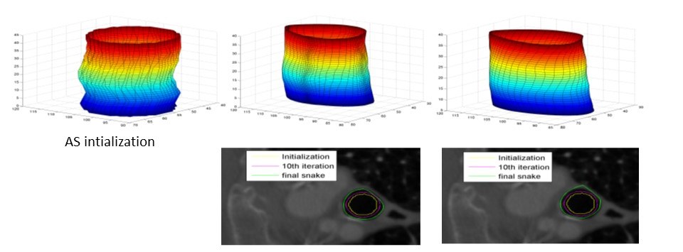
Volumetric scans such as computed tomography describe anatomy in three dimensions, but extracting a structure of interest slice by slice is slow and error-prone. We develop deformable models that segment 3D structures directly.
Our approach combines generalised cylinders with active surfaces and active contours (“snakes”) that start from a simple shape and iteratively adapt to the boundaries in the image, for example along tubular structures such as blood vessels. Related annotation tools help experts prepare reference data for computer-aided diagnosis.
- Generalised cylinders and active surfaces
- Deformable contour models
- Image annotation for computer-aided diagnosis
Related publications
Selected work from the lab in this area.
Novel Cost Function Definition for Minimum-Cost Tractography in MR Diffusion Tensor Imaging
In GeNeDis 2018. Springer International Publishing, pp. 135–150.
A Tractography Algorithm for MR Diffusion Tensor Imaging Based on Minimum-Cost Path
In XIV Mediterranean Conference on Medical and Biological Engineering and Computing 2016. Springer International Publishing, pp. 302–307.
Ratsnake: A Versatile Image Annotation Tool with Application to Computer-Aided Diagnosis
The Scientific World Journal, 2014, pp. 1–12.
A novel tool for segmenting 3D medical images based on generalized cylinders and active surfaces
Computer Methods and Programs in Biomedicine, 111(1), pp. 148–165.
An Intelligent Tool for Anatomical Object Segmentation Using Deformable Surfaces
In Artificial Intelligence: Theories and Applications. Springer Berlin Heidelberg, pp. 206–213.
Automated Segmentation and Registration of Dermatological Images
Journal of Mathematical Modelling and Algorithms, 2(3), pp. 277–294.